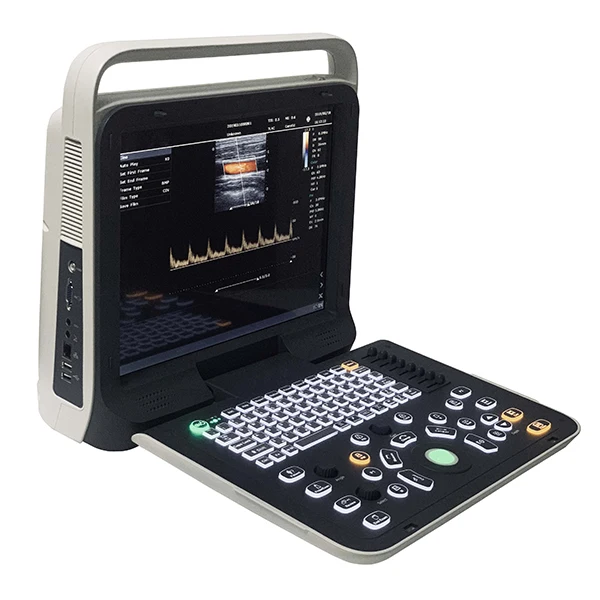
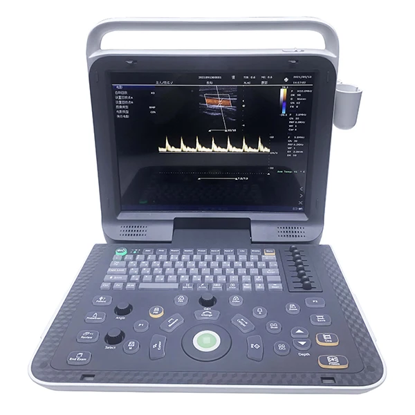
C10B Color Doppler Ultrasound Machine Portable Device
- Category: >>>
- Supplier: Guangzhou Sonostar Technologies Co. Limited
Share on (1600848733240):
Product Overview
Description
C10B


1 General Information
A brand_new ultrasound diagnostic platform with Innovations in areas of digital electronics achieve a new level of ultrasound diagnostic precision and higher diagnostic confidence.
A revolutionary workflow control is provided with the user-centric architecture of the new software platform.
2 Main technical parameters and functions
2.1 Technical platform
linux +ARM+FPGA
2.2 Channels and elements
Number of physical channels: ≥64
Number of probe array element number:≥128
2.3 Size and weight
Machine size:37cm(width) *36cm(height) * 15cm(thickness)
Package size: 46cm ×46cm × 31cm
Machine weight : 8.4kg
Weight including package :12kg(machine + 2 probes + packing)
2.4 Monitor
15-inch, high resolution, progressive scan, Wide Angle of view
Resolution:1024*768 pixels
Image display area is 640*480
2.5 Hard disk
Internal 500GB hard disk for patient database management
Allow storage of patient studies that include images,clips,reports and measurements
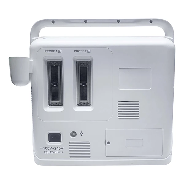
2.6 Transducer Ports
Two active universal transducer ports that support standard(curved array, linear array), high-density Probe
156-pin connection
Unique industrial design provides easy access to all transducer ports
2.7 Probe available
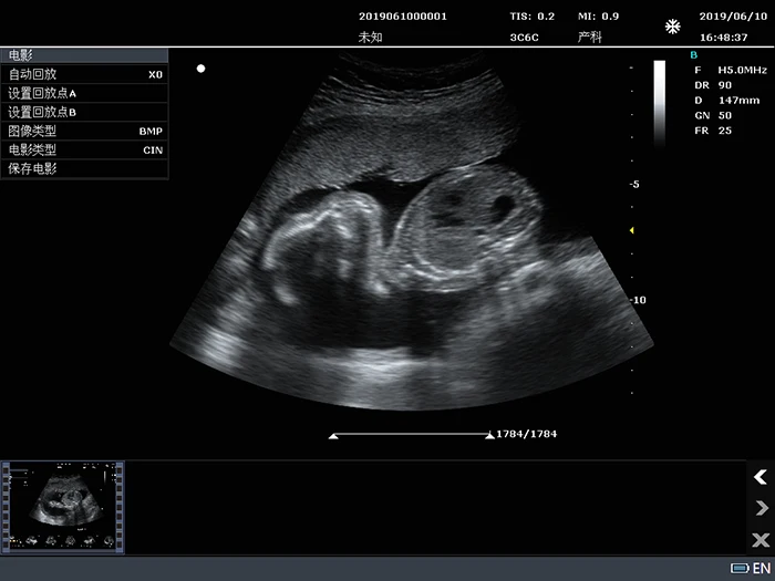
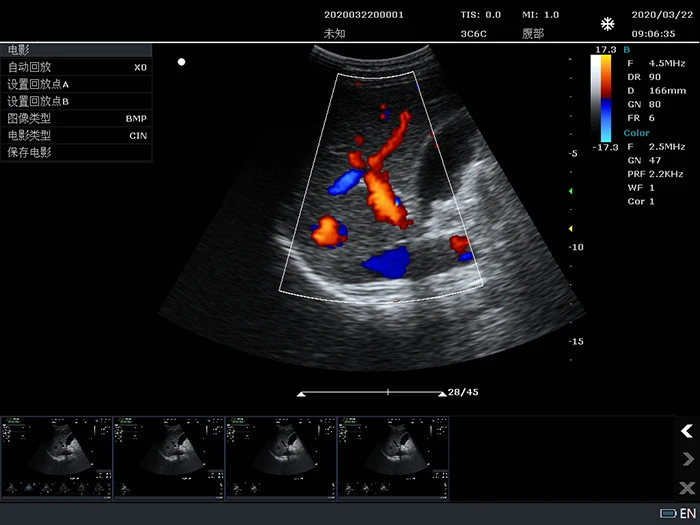
3C6C: 3.5MHz/R60/128,Convex array probe
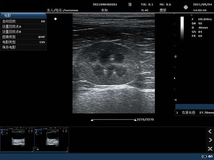
7L4C: 7.5MHz/L38mm/128,Convex array probe
10L25C: 10MHz/25mm/128,Convex array probe
6E1C: 6.5MHz/R10/128,EndocavityConvex array probe;
6C15C: 6.5MHz/R15/128,Micro convex array probe;;
3C20C: 3.5MHz/R20/128,Micro convex array probe;
6E1C: 6.5MHz/R10/128,EndocavityConvex array probe for Visual abortion;
6I7C: 6MHz/L64mm/128,IntrarectalLineararray probe;
2P2F:2.7MHz/L16mm/64 Phased arrayprobe;
5P2F:5.0MHz/L10mm/64 Phased arrayprobe;
2.8 Imaging modes
B-mode: Fundamental and Tissue harmonic imaging
Color Flow Mapping (Color)
B/BC Dual Real-Time
Power Doppler Imaging (PDI)
PW Doppler
M-mode
2.9 frequency number
B/M:Fundamental wave,≥3;harmonic wave: ≥2
Color/PDI:≥2
PW: ≥2
2.10 Cine
B mode: ≥5000frames
B+Color/B+PDI mode: ≥2300frames
M,PW: ≥ 190s
2.11 image zoom
available on live, 2B, 4B and reviewed images
up to 10X zoom
2.12 image save
format:
BMP,JPG,FRM(single image);
CIN,AVI(multiple images)
Support DICOM, conform to DICOM3.0 standard
Built in workstation,support patient data search and browse
4 Exam Modes
Abdomen
Obstetrics
Gynecology
Fetal Heart
Small parts
Urology
Carotid
Thyroid
Breast
Vascular
Kidney
Pediatrics
3. Product configuration
5 Standard configuration
Host(Built-in 500G hard disk)
3C6C convex array probe
7L4C linear array probe
User's Manual
Power cable


Certifications

Exhibition

We Recommend

New Arrivals
New products from manufacturers at wholesale prices