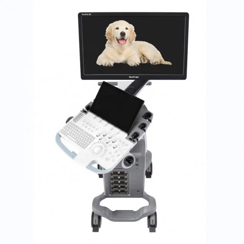
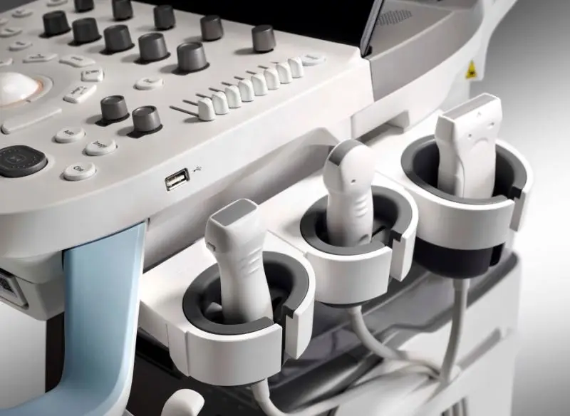
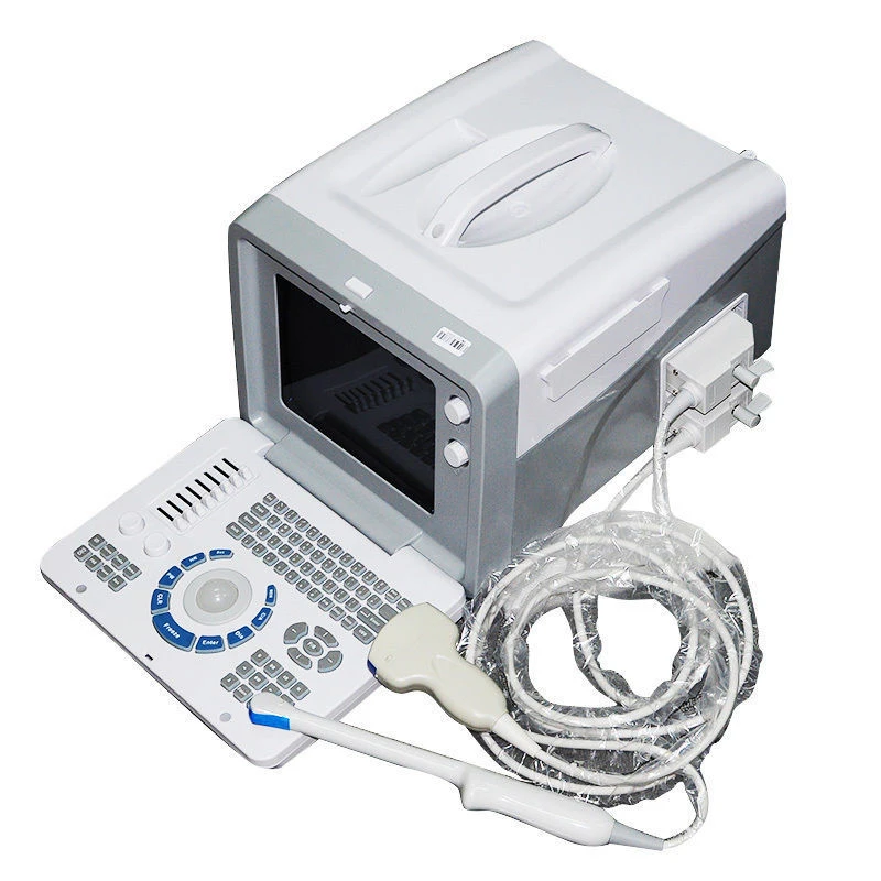
4D/5D Ultrasound Machine Veterinary/17Mhz High Frequency Ultrasound SonoScape Ultrasound Imaging Diagnosis
- Category: >>>
- Supplier: Medsinglong Co. Ltd.Medsinglong Ltd.
Share on (1601258373585):
Product Overview
Description
Product Description

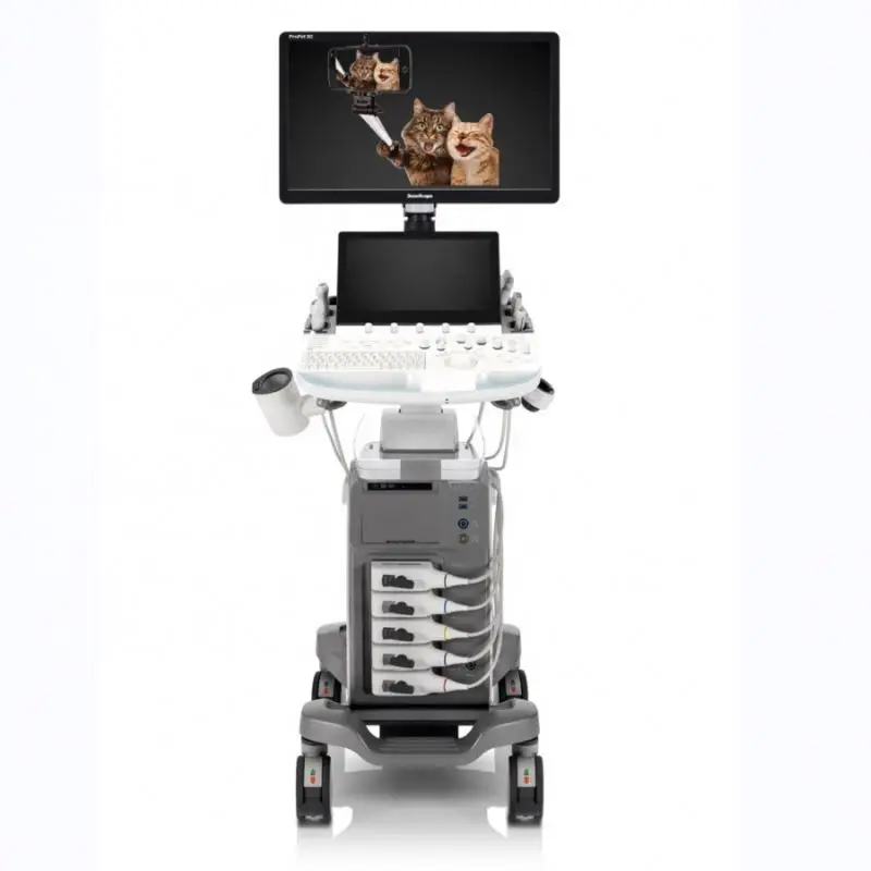
All creatures are great and small

Sono Assistant
Guides clinicians through the entire exam and provides customizable scanning protocol helping streamline workflow while increasing
standardization.
standardization.

Animal’s name, species, breed and castration status
Saves complicated input time
Saves complicated input time
Strain Elastography
Offers a real-time tissue stiffness assessment displayed as a color map to detect potential abnormalities within normal tissue.
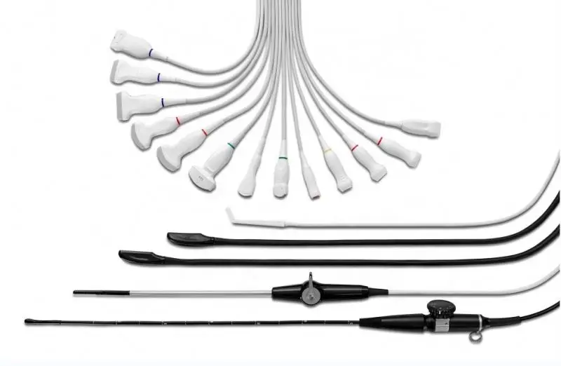
Available on linear, convex, micro-convex, and endo-cavity transducers to cover a wide range of applications.
Available on linear, convex, micro-convex, and endo-cavity transducers to cover a wide range of applications.
Contrast Enhanced Ultrasound
The non-linear contrast enhanced ultrasound imaging makes full use of harmonic and fundamental signals to give a more enhanced image of difficult-to-view blood flow.
Vis-Needle
Reveals needle location within animal anatomy with no distortion when performing interventions like nerve blocks and tissue biopsies.
Micro F
Distinguishes minute flow from overlaying tissue movement effectively and depicts hemodynamic with higher sensitivity and spatial resolution.
μ-Scan+
Is delicately engineered to reduce speckles while improve image uniformity and enhance border continuity, providing authentic presentation of details and enhanced lesion display.
HQ Scan
HQ scan is a newly-developed 2D improvement technology to enhance the contrast and whole image structure with better details. It is more helpful to visualize the border and margin of space-occupying lesions, nodules and tumors in parenchymatous organs.

TDI
TDI uses myocardial Doppler frequency shifts to quantify myocardial tissue motion, with red and blue representing the different
directions of wall movement. Combining TDI with PW is to better obtain the motion trajectory of the myocardial wall.

MQA
Precise left ventricular wall motion detection with globally 2D speckle patterns tracking provides accurate quantitative analysis
including strain, strain rate, displacement, velocity, etc. on myocardial walls.
including strain, strain rate, displacement, velocity, etc. on myocardial walls.
Stress Echo
Takes multiple dynamic images at rest and after stress and makes side by side comparison. Professional wall motion bulls-eye scoring and reporting is provided for further effective evaluation of animal cardiac muscle viability.
LVO
Enables to enhance discrimination between myocardial tissue and blood pool, providing better visualization of the endocardia border display especially for difficult patients.
Auto EF
Save more time and efforts compared with manual measurement with Auto EF, which calculates ejection fraction based on left ventricular wall tracing and Simpson’s rule.
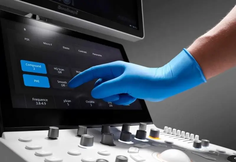
Auto Optimization
Provides intuitive one touch 2D, Color and Dopple image optimization through intelligent real-time algorithms.
Auto Bladder
One key bladder wall tracing and volume measurement from Auto Bladder can efficiently provide more accurate contour and results, which is not subject to the bladder shape and size.
Auto EF
Based on left ventricular wall tracing and Simpson's rule saves time and efforts compared with manual measurement.
Auto Trace
It can trace the PW/CW spectrum automatically, which help doctors make measurement easily and conveniently.
Clinical Images

Item Name | 4D/5D Ultrasound Machine Veterinary/17Mhz High Frequency Ultrasound SonoScape Ultrasoud Imaging Diagnosis |
Introduction | ProPet 80, the premium veterinary ultrasound imaging system, adopts the most cutting-edge ultrasound technology to deliver quick diagnostic information, advanced functionality and an ergonomic system to tackle the rising number of challenging cases and ever-increasing diversified demands for veterinary practice on all fronts. It is indeed worth considering if you need a hospital-based machine capable of high end ultrasonography. |
Imaging Modes & Imaging Technology | 2B & 4B Mode |
PHI/THI | |
M Mode | |
CFM | |
PDI / DPDI | |
PW | |
CW | |
HPRF | |
LGC | |
TSI | |
Compound | |
μ-Scan | |
HQ Scan | |
SR Flow | |
Bright Flow | |
Micro F | |
Function | Panoramic Imaging |
Widescan | |
Elasto | |
Contrast Imaging | |
Rotation | |
Biopsy | |
Vis-Needle | |
Auto Trace | |
TEI | |
TDI | |
Color M | |
AMM | |
Auto EF | |
G-MQA | |
R-MQA | |
Stress Echo | |
LVO | |
MCE(Only for Tender) | |
Auto Bladder | |
Freehand 3D | |
Other features | DICOM3.0 |
Sono Drop | |
Auto Optimization | |
Sono Assistant | |
Sono Synch |
Company Profile


FAQ
1. Who are we?
We are based in Guangdong, China, start from 2013,sell to Southeast Asia(18.00%),Africa(18.00%),North America(15.00%),South America(12.00%),Mid East(8.00%),Southern Europe(8.00%),Western Europe(6.00%),Central America(3.00%),South Asia(3.00%),Eastern Europe(2.00%),Oceania(2.00%),Northern Europe(2.00%),Domestic Market(2.00%),Eastern Asia(00.00%). There are total about 51-100 people in our office.
2. How can we guarantee quality?
Always a pre-production sample before mass production;
Always final Inspection before shipment;Have our own engineer team who speak English very good stand by 24 hours a day, 7 days a week for maintenance.
The only company who can offer u refund money in case of bad quality product
3.What can you buy from us?
Medical Image,Ultrasound Machine, Panel Detector, X-Ray Machine, Lab Analyzers,IVD,POCT,Medical Consumables,Beauty Equipment
4. Why should you buy from us not from other suppliers?
1) 8%-30% competitive price than others 2) We can offer one-stop shopping for our customer for buy medical equipment for hospital
3). 12-36 months After sale service warranty
5. What services can we provide?
Accepted Delivery Terms: FOB,CFR,CIF,EXW,FAS,Express Delivery;
Accepted Payment Currency:USD,EUR,JPY,CAD,AUD,HKD,GBP,CNY,CHF;
Accepted Payment Type: T/T,L/C,MoneyGram,Credit Card,PayPal,Western Union,Cash;
Language Spoken:English,Chinese,Spanish,French
We Recommend
New Arrivals
New products from manufacturers at wholesale prices