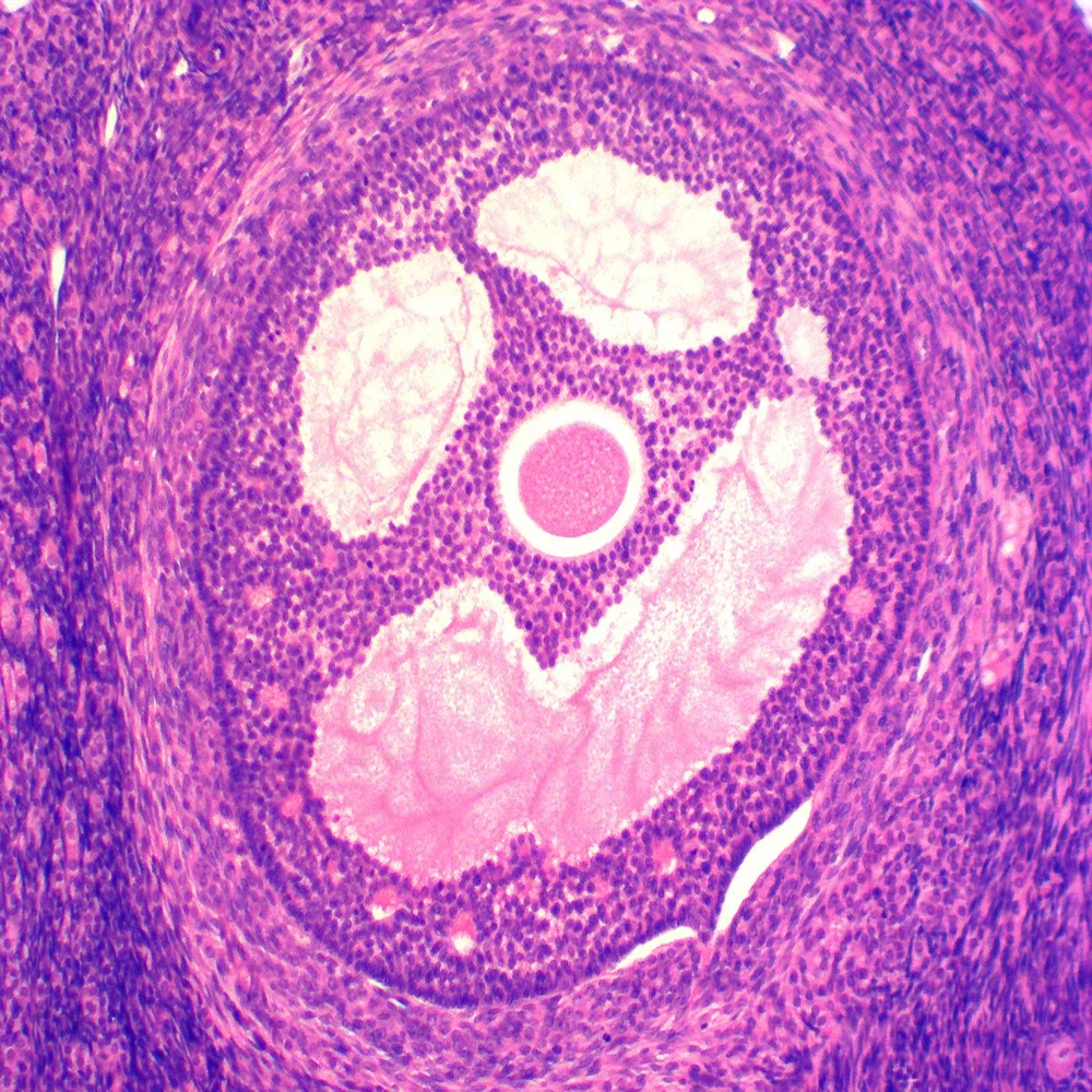
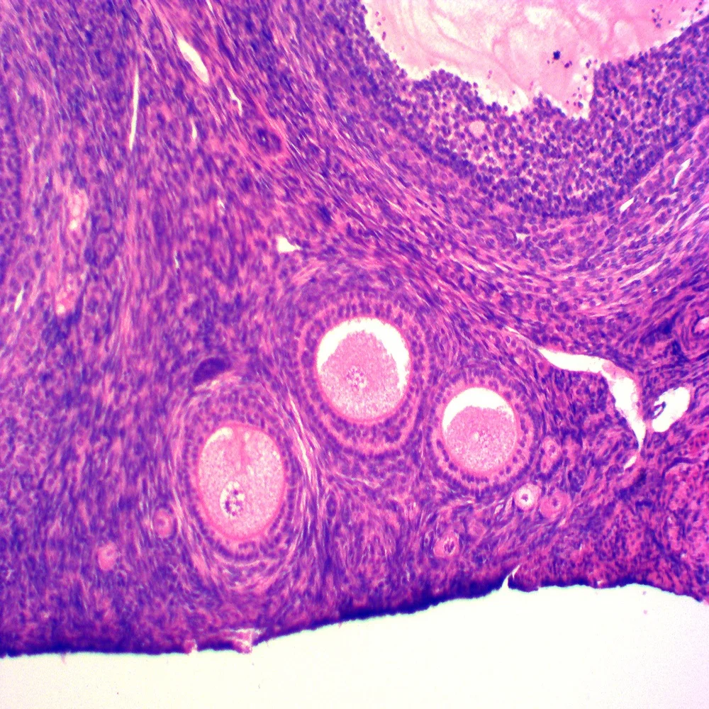
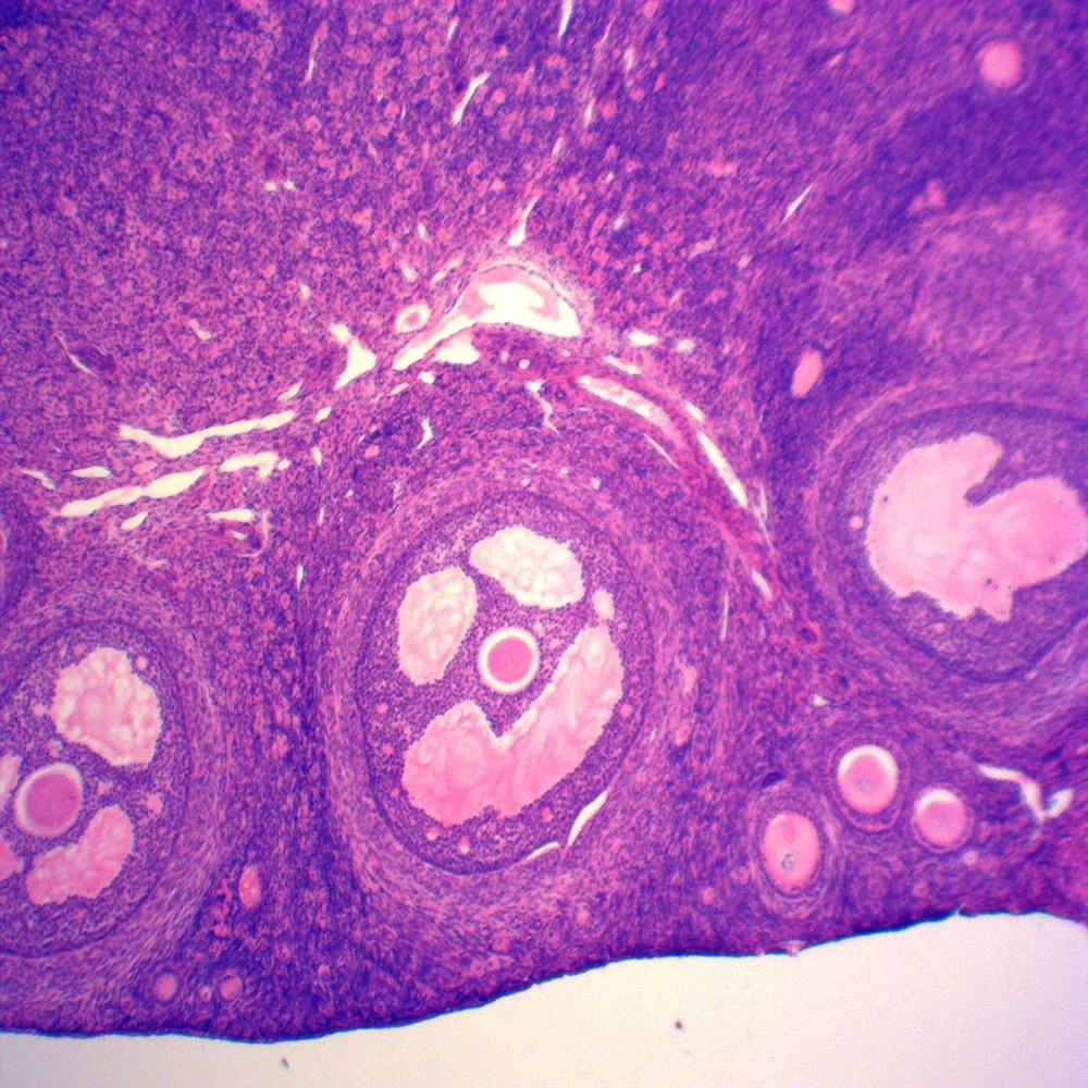
Teaching resources Medical science histology prepared microscope slides/Ovary of Rabbit sec histology prepared slides
- Category: Other Teaching Resources >>>
- Supplier: Henan Yulin Edu. Project Co. Ltd.
Share on (391248732):
Product Overview
Description
Teaching resources Medical science histology prepared microscope slides/Ovary of Rabbit sec histology prepared slides
Microscope Prepared Slies List
Botany
| YBB010010 | Epidermis of Allium cepa W.M. |
| YBB010020 | Epidermis cells of Capsicum frutescens Fruit W.M.(show simple pit-pair) |
| YBB010030 | Plasmodesmus sec.(endospermium of Diospyros kaki ) |
| YBB010040 | Root tip of Allium cepa L.S.(show mitotic division) |
| YBB010050 | Chromosome of Allium W.M. |
| YBB010060 | Stem apex of Hydrilla verticillata L.S.(show apical meristem) |
| YBB010070 | Syringa oblata bud L.S. |
Zoology
| YZO010010 | Euglena W.M. |
| YZO010020 | Amoeba W.M. |
| YZO010030 | Paramecium caudatum W.M. |
| YZO010040 | Conjugation of Paramecium caudatum W.M. |
| YZO010050 | Binary fission of Paramecium caudatum W.M. |
| YZO010060 | Vorticella W.M. |
| YZO010070 | Stylonychia W.M. |
| YZO010080 | Epiphanes sepia W.M. |
Parasitology
| YMP060150 | Immature proglottid of Dipylidium caninum W.M. |
| YMP060160 | Scolex of Dipylidium caninumW.M. |
| YMP060170 | Gravid proglottid of Dipylidium caninum W.M. |
| YMP060180 | proglottis of Moniezea expansa T.S. |
| YMP060230 | Scolex of Helictometra W.M. |
| YMP060240 | Gravid proglottid of Helictometra W.M. |
| YMP060250 | Mature proglottid of Helictometra W.M. |
| YZO060010 | Ascaris lumbricoides (male )T.S. |
| YZO060020 | Ascaris lumbricoides (female) T.S. |
| YZO060030 | Ascaris lumbricoides (female and male)T.S. |
1.Different kinds of slides
Histology section and Embryology section: Epithelial tissue section. Connective Tissue section. Muscle tissue section. Nervous system tissue section. Circulatory system tissue section. Immunologic system tissue section. Endocrine system tissue section. Digestive system tissue section. Respiratory system tissue section. Urinary system tissue section. Genital system tissue section. Sense organ tissue section. Embryon tissue section. Section of cytobiology and genetics.
1)Extract from fresh material. Keep the whole structure.
2)Clearly cell tissue, keep the original shape.
3)Well-distributed coloration: HE, India ink stain, alcohol magenta, hematoxylin staining.
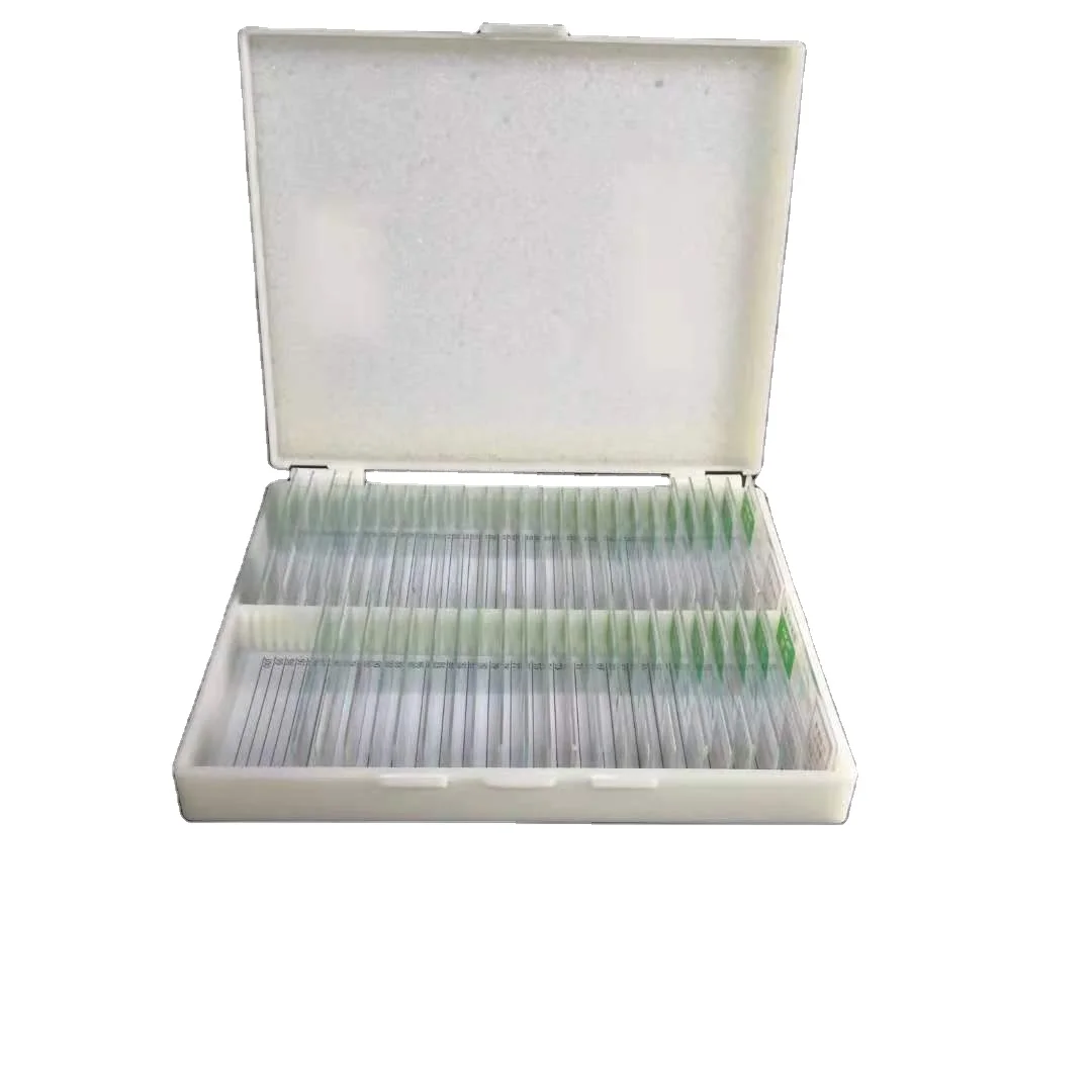
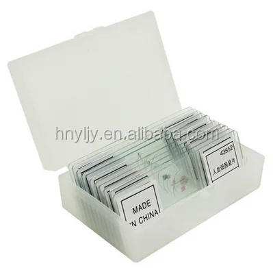
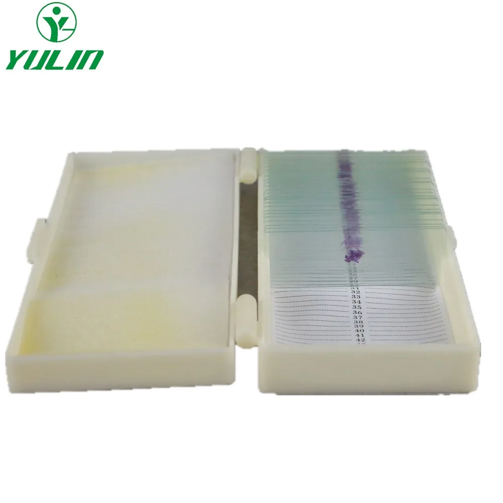
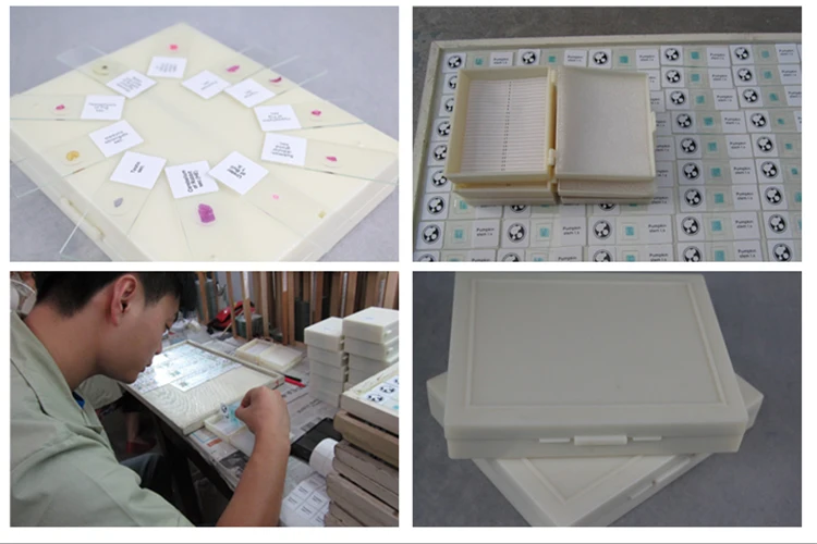
4)Packing as you like, plastic or wooden slides box.
5)Easy to stimulate students' interest in study the zoology and structure of animal will be helpful to improve the experiment quality and teaching effect.
6)ISO9001:2008, ISO14001-2004 standard, GBT28001-2001 standard, AA credit grade, attend educational association, China education equipment industry association, Henan educational instrument and equipment association.
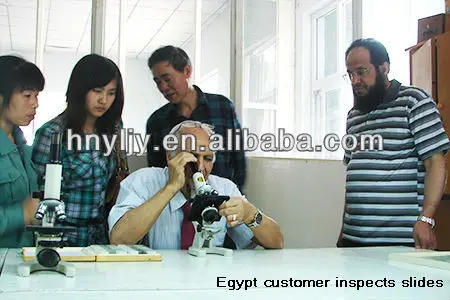
7)Exported more than 23 countries, such as Germany, Japan, UK, France, Belgium, Australia, New Zealand, Indonesia, Egypt, Syria, Iran, Malaysia, Thailand, Hong Kong, Spain, Turkey, ect.
2.Microscope slides detail information:
1)Staining method: Hematoxylin and eosin staining, silver staining, gold chloride staining, COX staining;
2)Thickness of section: 3μm~5μm.
3)Customers: all levels of students/home school programs
4)Well-distributed coloration:Hematoxylin and eosin staining, silver staining
5)Material: microscope glass slides, durable and directly view
6)Size: 76.2x25.4x1-1.2mm(3''x1'') length/width/thickness
7)Weight: 100PCS plastic box N/W 0.2KG, G/W 0.75KG
100PCS wooden box N/W 0.25KG,G/W 0.8KG
Transparent label: Glossy paper for stickers; good label English
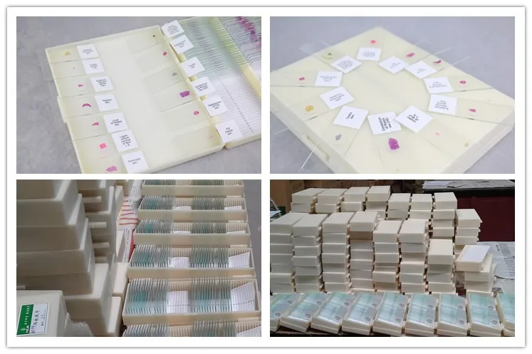
8)Packing: packed in wooden or plastic box; 100/50/26/25/15 pieces plastic or wooden slide box; neutral bleached paper carton(61*38*31CM); there has label with introduction in English on slide and cartons.
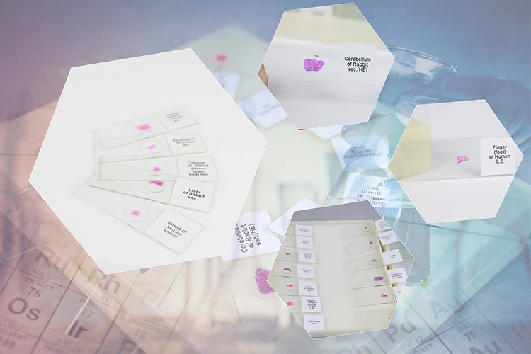
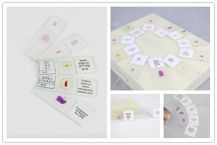
Slides Pictures




3.About Us
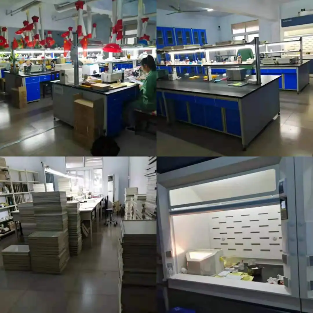
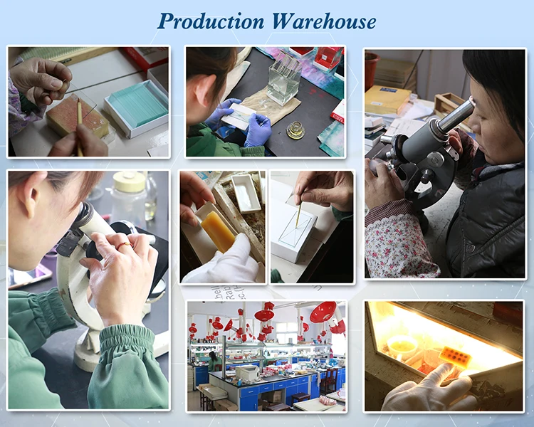
We are a professional manufacturer of science and educational supplier for primary schools, middle schools, colleges, hospitals or medical institutes. Originating from a biology lab of the Henan Normal University in 1958, our company specializes in manufacturing microscope slides, microscope slides box. Different kinds of slides including botany, zoology, parasitology, histology, human pathology, microbiology, embryology slides, ect. The slide was cut subtly without any mark, break or constrict. There is no destruction of tissues or cells.The spread of tissues has clear bounds; they remain the original shape. Also the coloration for the tissues is obvious and clear. As more than 53 years professional manufacture, we have an excellent reputation from domestic and overseas. We have exported more than 23 countries, such as Germany, Japan, UK, France, Belgium, Australia, New Zealand, Indonesia, Egypt, Syria, Iran, Malaysia, Thailand, Hong Kong, Spain, Turkey, ect. They all praised our slides quality is best .


We are a factory of all kinds of prepared microscope slides , microscopes , microscope slides and cover slips , teaching and medical models , and many other kinds of educational products , any demands you have , please contact me freely .
Waiting for your early reply
Best Regards
Henan Yulin Edu. Project Co., Ltd.
Cathy Wang Export manager
Email: cathywang @ hnylju.cn or sales @ yulinedu1958.com
Wechat: 18637306387
trademanager: hnyljy
Skype: jessy-zhang0715
Phone: 0086-13503800666
QQ: 2469484693
Website: //www.yulinedu1958.com/
We Recommend


New Arrivals
New products from manufacturers at wholesale prices