* PC: Intel i3 3.0GHz,1T hard disc,4G memory,23″1920×1080 LCD, windows 7.
* 16 grades noise reduction, vertical and horizontal mirror image, positive and negative image, last frame hold, gamma correction, auto bright and contrast correction, DICOM3.0,medical record management, and so on.
(7) X-Rray Field Localizer: 4 laser dots.
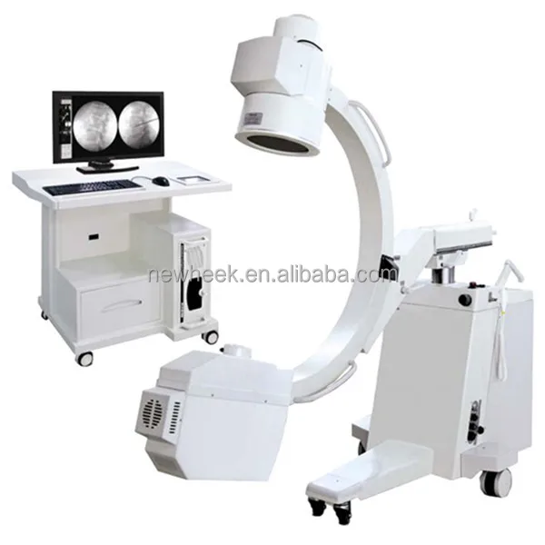
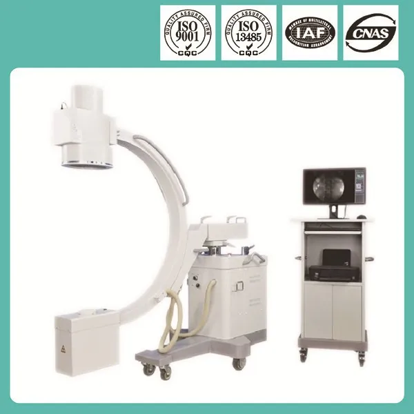
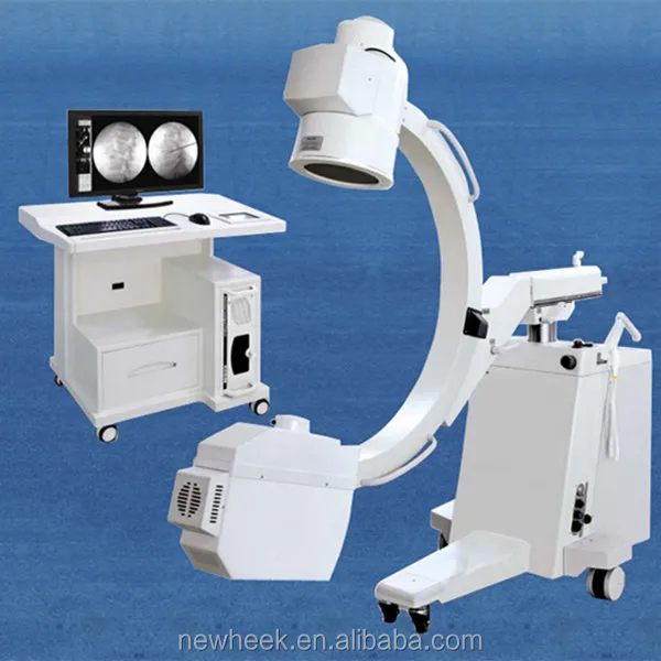
(8) C-arm Mechanical Specification
Outer diameter of c-arm
1.3m
Source to image SID
1030mm
Operation space
≥870mm
C-arm vertical movement
≥400mm, motor drive
C-arm rotation
-180° - +180° , manual
C-arm slide angle
-25° - +90° , manual
C-arm front and back movement
≥200mm, manual
C-arm horizontal swing
-12.5° - +12.5°, manual
Steering shank with brake
-90° - +90°, manual
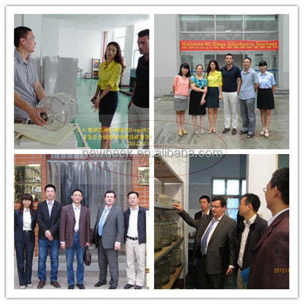
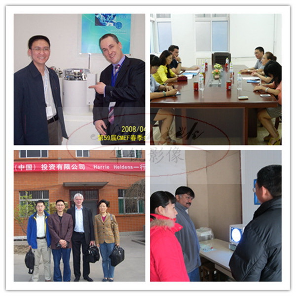
Trade Shows
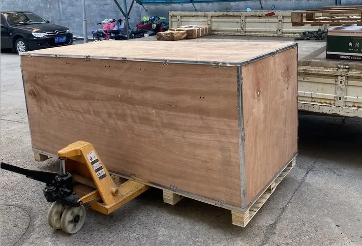
Packaging & Shipping
FAQ
FAQ -- Technical Knowledge of Mobile C-Arm X-Ray Machine
1. Q: What is the main components of c-arm?
A: It includes machine case, display, invertor, integrated generator and tube, image intensifier, image process system, console and C-frame etc.
2. Q: How does c-arm work?
A: Start the c-arm to the projection position, make the image intensifier as near as to the patient projection body part, which could obtain the sharp image. Press the foot switch,
the x-ray come out from the integrated tube and pass though the body part, the image intensifier captures the x-ray then change them into electric signal, then the electric signal come into image process system and after being processed displayed on the monitor. The fluoroscopy work finish. The doctor can see the fluoroscopy density image on the monitor.
3. How to maintain the c-arm?
A: (1) The mechanical part (such as lifting and c-arm sliding) of mobile c-arm needs to keep dampproof.
(2) Don’t squeez the cables of between the console and case during c-arm moving.
(3) When the machine deesn’t work, it is better to let the intergrated generator and tube down and let the image intensifier up. Becuase the grid is in the front of image intensifier and it easy to damage when collide.
(4) Don’t put magenetic project on or near image intensifier to avoide the image intensifier magnetization.
(5) Please keep the doctor with protection method during operation.
(6) Keep the machine room clear and dry and no sundries.
(7) Regular check the c-arm components fixed connection, make clearance and apply oil (don't make the high voltage cables touch oil), when nessary, it needs to make rectify all the parameters by instrument. If the machine doesn't use for a long time, it needs to make careful check and rectify before using.