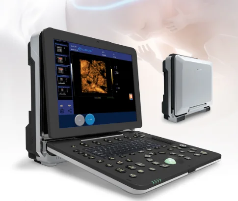
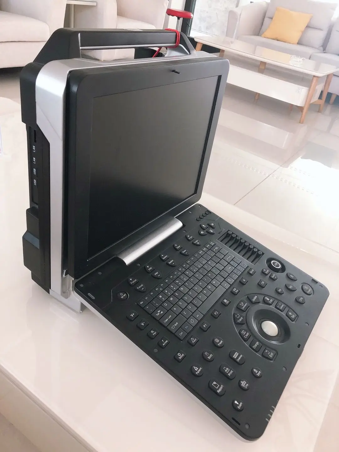
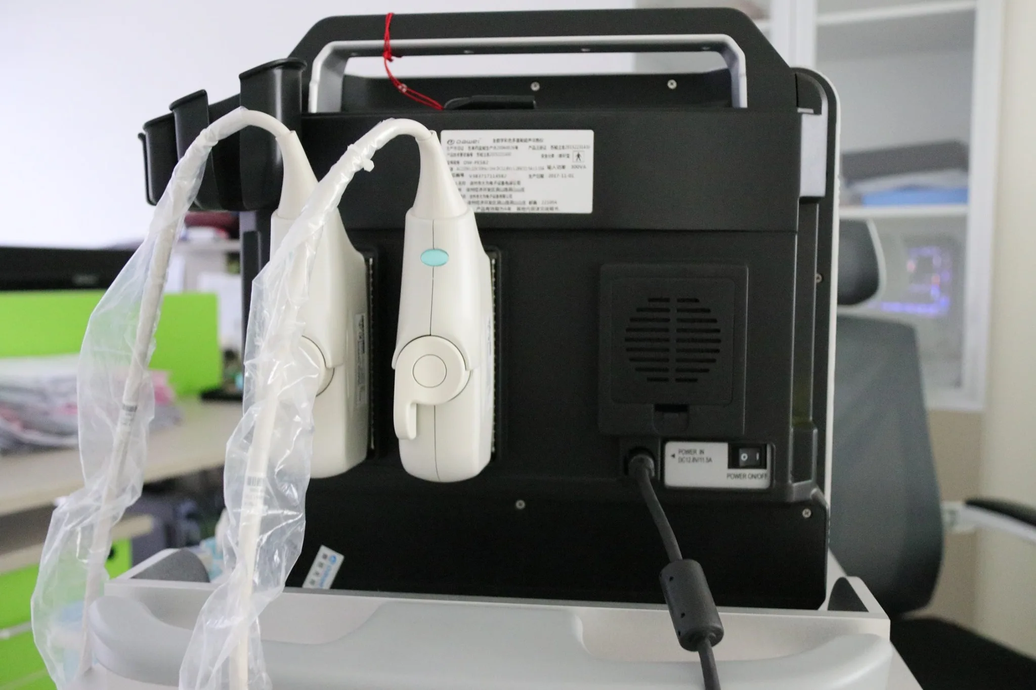
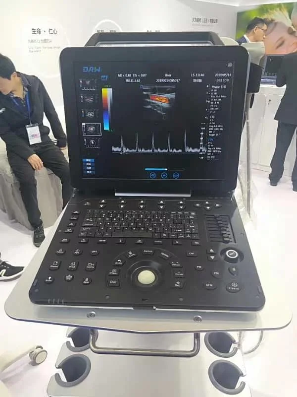
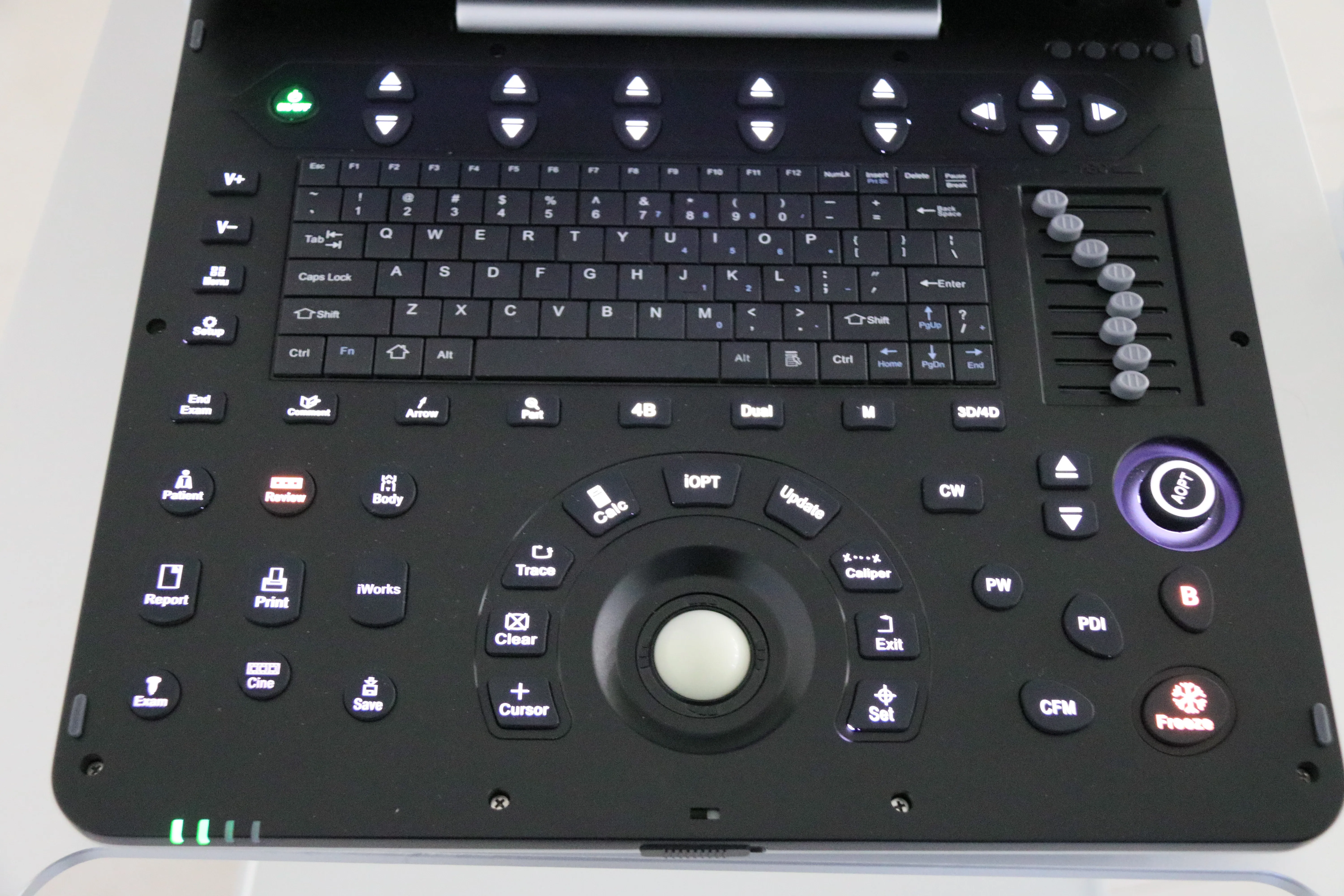
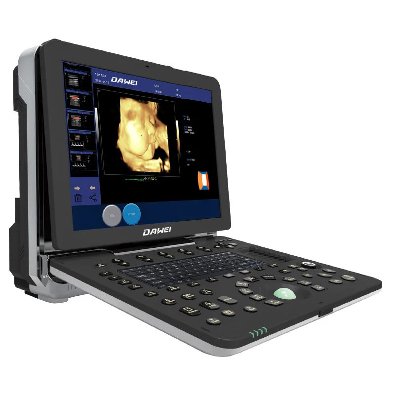
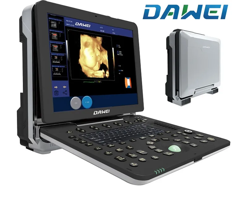
DW P6 4D портативный УЗИ ультразвуковой
- Категория: >>>
- Поставщик: Dawei Medical (jiangsu) Co. Ltd.
Сохранить в закладки 60757995704:
Описание и отзывы
Характеристики

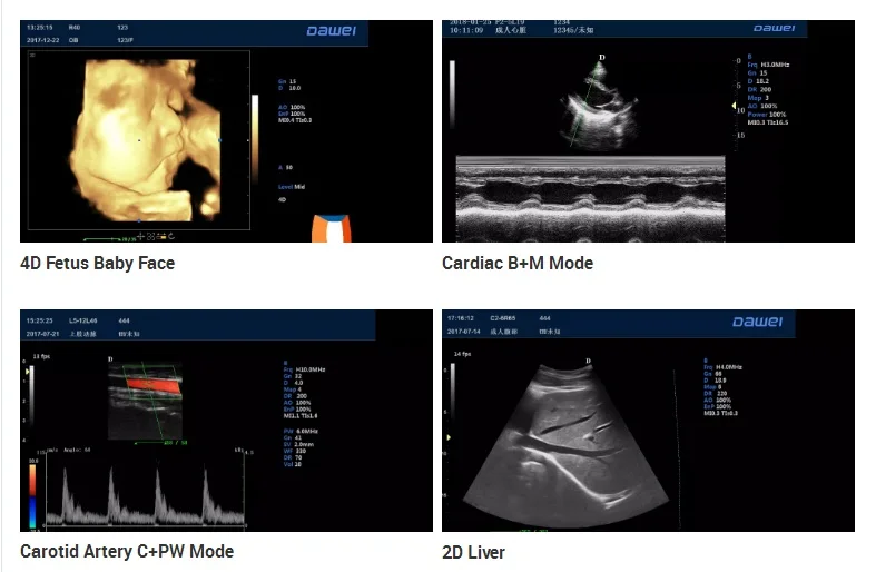
DW-P6 Portable doppler ultrasound system and 4D color doppler
Main Specification:
1)Full-Digital 2D gray scale imaging
2)Full-Digital Tissue Harmonic Imaging (THI)
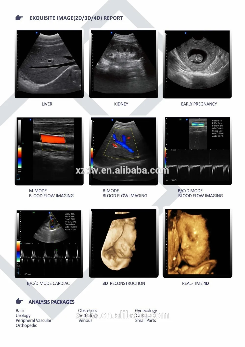
3)Color Doppler blood flow imaging
4)Directional color energy Doppler imaging
5)Pulse Wave Doppler imaging (PW)
6)Continuous Wave Doppler imaging (CW)
7)Space compound imaging
8)Trapezoidal imaging
9)2D, color, Doppler mode automatic optimization adjustment technology
10)Real-time triple synchronizing
11)Adaptive speckle suppression technology
12)Real-time 3D imaging
13)Intelligent picture - in - picture imaging mode (PIP)
14)Monitor: 15 inch high resolution medical LCD monitor, adjustable angle
15)Probe connectors: ≥2 active
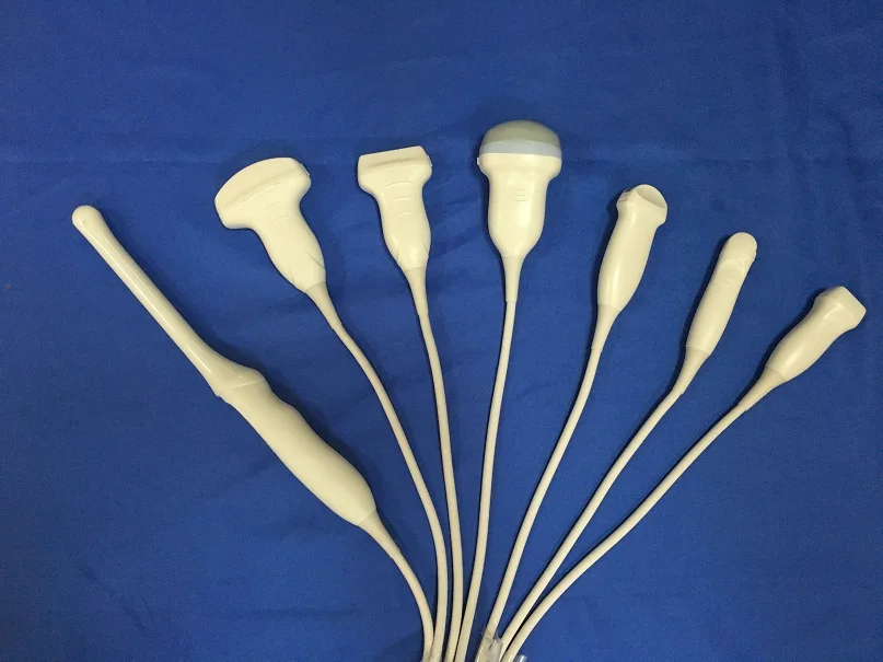
1 Multiple probe configuration:
1.1 Convex probe frequency: 2.0-5.0MHZ(multi-frequency, Harmonic frequency≥5 ), probe scanning angle 20°~85°, visible and adjustable.
1.2 Linear probe frequency: 6.0-12.0MHZ(multi-frequency, harmonic frequency ≥4 ), probe scanning with trapezoidal imaging technology and 2D beam deflection technology
1.3 Trans-vaginal probe frequency: 5.0-9.0MHZ(multi-frequency, harmonic frequency ≥2 ), probe scanning angle 20°~160°, visible and adjustable.
1.4 Real time 3D (4D/5D) volume probe frequency: 2.0-6.0MHz, 4 segments multi-frequency.
1.5 Phased array probe frequency: 2.5-4.0MHz, 3 segments multi-frequency.
2 2D imaging mode
2.1 Gray scale: 256
2.2 Gray Map: ≥16 level, visible and adjustable
2.3 Dynamic range: 20-280db(visible and adjustable)
2.4 Resolution: Horizontal≤1mm; Vertical≤0.5mm
2.5 Under B mode, focus number: 1-6, focus position continuously adjustable
2.6 STC gain control ≥8 segments
2.7 THI: harmonic frequency ≥2 segments
2.8 Line density: ≥256, visible and adjustable
2.9 Preset: ≥40 kinds, users can customize the inspection conditions for the optimized images of different organs
2.10 Max scanning depth: ≥31cm, visible and adjustable
2.11 Scanning angle: 50°-100°, visible and adjustable
2.12 Cine loop ≥4800 frames
2.13 Adaptive speckle suppressio: 0-100 adjustable
2.14 Amplification: overall amplification, local amplification, M-type amplification(do M-type sampling amplification under both scanning or freeze state)
3 Color blood flow imaging mode:
3.1 Color gain: adjustable
3.2 Color frequency: ≥3 kinds, visible and adjustable
3.3 Sampling frame: size and position adjustable
3.4 Color blood flow steer: ≥3 angles adjustable
3.5 Color map: 1—9 level
3.6 Color persist: 0—6 level
3.7 B/C split display: available
Applications: abdominal, urology, OB&GYN, paediatrics / neonatal, superficial / small organ, musculoskeletal, cardiology etc.
Convex probe- Adult abdomen application
Frequency: 2.5, 3, 3.5, 4, H4, H5MHz
Power:5-100% (arithmetic progression of 5: 5,10,15...100)
Gain:0-100
Dynamic range: 20-280% (geometric progression of 2 start from 20: 20,40,60...280)
Gray map:0-7
Frame correlation:0-4
Filtering:0-4
Image denoising:0-14
Scanning depth:3-27.3cm
Body mark:17
Scanning range:50-100% (arithmetic progression of 10 start from 50: 50,60,70...100)
Focus point:6
Pseudo color map :0-11
Linear density:64,128,256
TSI: normal, fat, fluid, muscle
Reversal:up/down, left/right
Compound frequency: on/off
Automatic optimization: on/off
Space compound: on/off
Linear probe-Carotid application
Frequency: 6, 7.5, 8.5, 10, H10 MHz
Power:5-100% (arithmetic progression of 5: 5,10,15...100)
Gain:0-100
Dynamic range: 20-280% (geometric progression of 2 start from 20: 20,40,60...280)
Gray map:0-7
Frame correlation:0-4
Filtering:0-4
Image denoising:0-14
Scanning depth:2-11cm
Body mark:13
Scanning range:50-100% (arithmetic progression of 10 start from 50: 50,60,70...100)
Focus point:5
Pseudo color map :0-11
Linear density:64,128,256
TSI: normal, fat, fluid, muscle
Reversal:up/down, left/right
Steering: left/right
Trapezoid Imaging: on/off
Compound frequency: on/off
Automatic optimization: on/off
Space compound: on/off
Cardiac probe- Adult cardiac application
Frequency: 2.5, 3, 3.5, 4, H3, H4 MHz
Power:5-100% (arithmetic progression of 5: 5,10,15...100)
Gain:0-100
Dynamic range: 20-280% (geometric progression of 2 start from 20: 20,40,60...280)
Gray map:0-7
Frame correlation:0-4
Filtering:0-4
Image denoising:0-14
Scanning depth:3-27.3cm
Body mark:7
Scanning range:50-100% (arithmetic progression of 10 start from 50: 50,60,70...100)
Focus point: 5
Pseudo color map :0-11
Linear density:64,128,256
TSI: normal, fat, fluid, muscle
Reversal:up/down, left/right
Compound frequency: on/off
Automatic optimization: on/off
Space compound: on/off
4D probe - Obstetrics applications
Frequency: 2, 3, 4.5, 6, H5 MHz
Power:5-100% (arithmetic progression of 5: 5,10,15...100)
Gain:0-100
Dynamic range: 20-280% (geometric progression of 2 start from 20: 20,40,60...280)
Gray map:0-7
Frame correlation:0-4
Filtering:0-4
Image denoising:0-14
Scanning depth:3-27.3cm
Body mark:7
Scanning range:50-100% (arithmetic progression of 10 start from 50: 50,60,70...100)
Focus point: 6
Pseudo color map :0-11
Linear density:64,128,256
TSI: normal, fat, fluid, muscle
Reversal:up/down, left/right
Compound frequency: on/off
Automatic optimization: on/off
Space compound: on/off
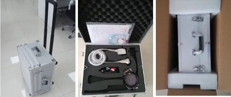
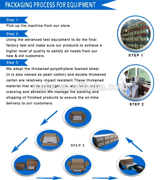
Packaging & Shipping

Company Information

Похожие товары
Брюки-карго лучшего качества, тактические брюки, осень для мужчин и женщин
12,00 $ - 14,00 $
Низкая цена балконные стеклянные перила с антикоррозийной балясиной из нержавеющей
45,00 $
Оптовая продажа, День матери, День Святого Валентина, свадьба, с наилучшими пожеланиями, роскошный подарок, музыкальный розовый медведь, Вечные розы из ПВХ, акриловая шкатулка для ювелирных изделий
1,20 $ - 6,00 $
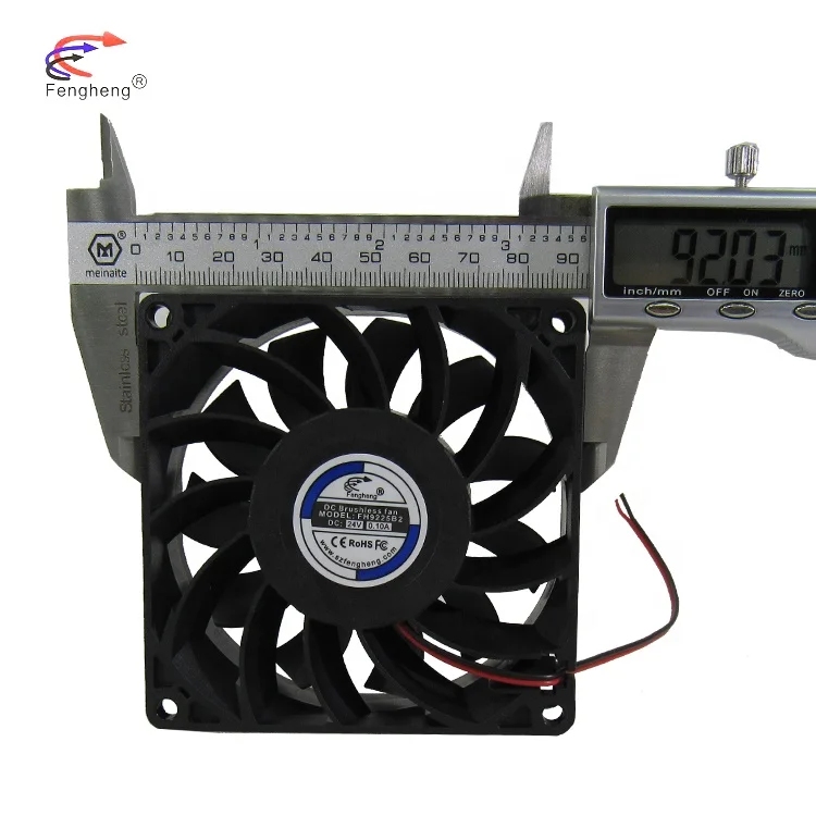
FH9225 вытяжной вентилятор для ноутбука 92x92x25 мм 12 В 24 в 48 в бесщеточные двигатели Вентилятор охлаждения постоянного тока
3,50 $ - 3,70 $
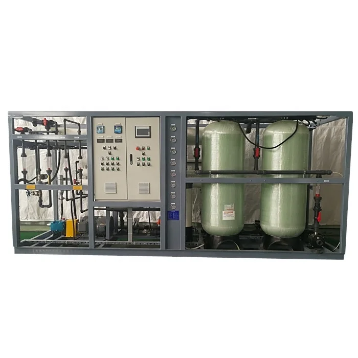
200m 3/день Смарт упакованы рекуперации энергии оборудования опреснения морской воды обратного осмоса Мембрана для безопасности питьевой воды
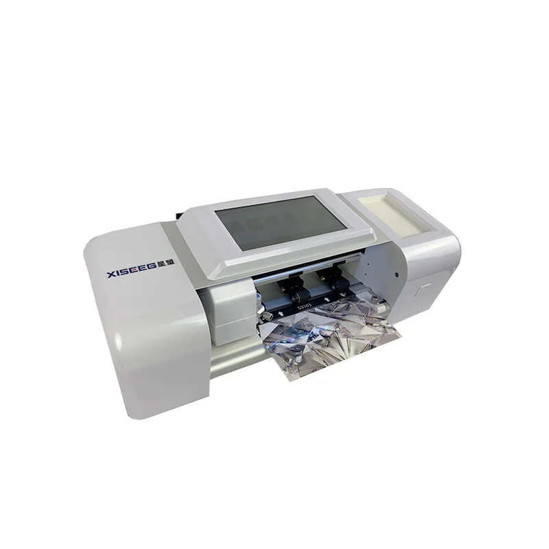
Мягкая пленка для телефона, режущая наклейка на кожу телефона, виниловый резак, защита экрана, гидрогелевый резак, машина для резки винилового принтера
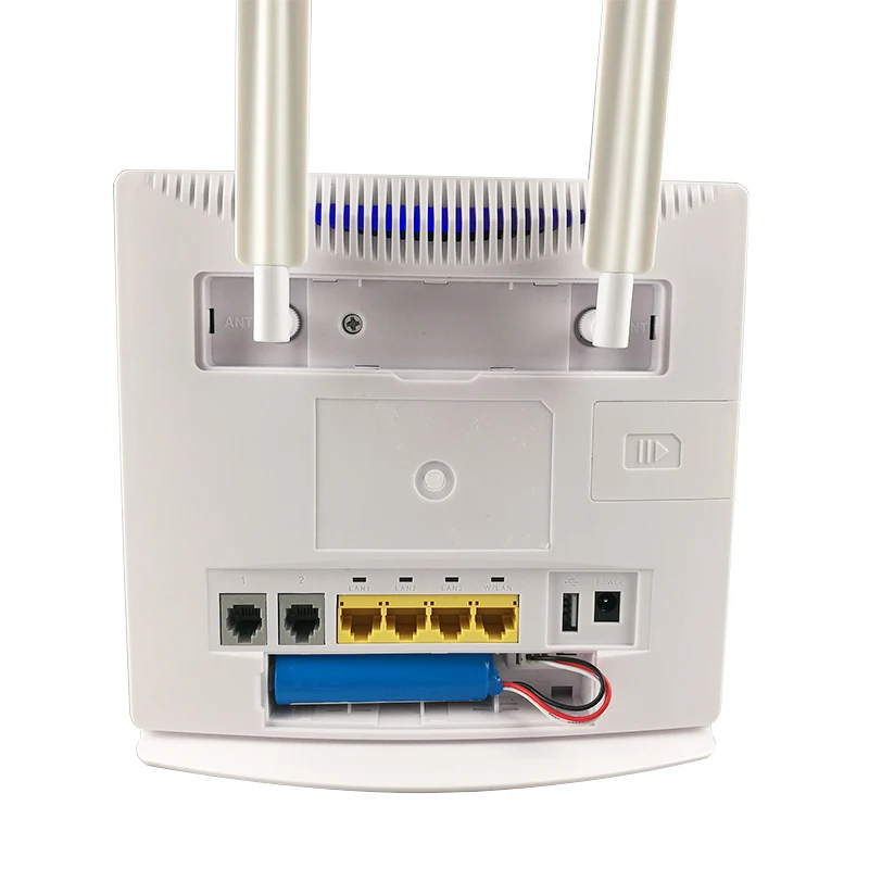
Роутер, идеально подходящий для поездок может использоваться как 2G, 3G, 4G, Telecom беспроводные маршрутизаторы ETS-B688 с SIM замок/изменить IMEI/точку доступа Wi-Fi
44,00 $ - 46,00 $
Новые поступления
Новинки товаров от производителей по оптовым ценам
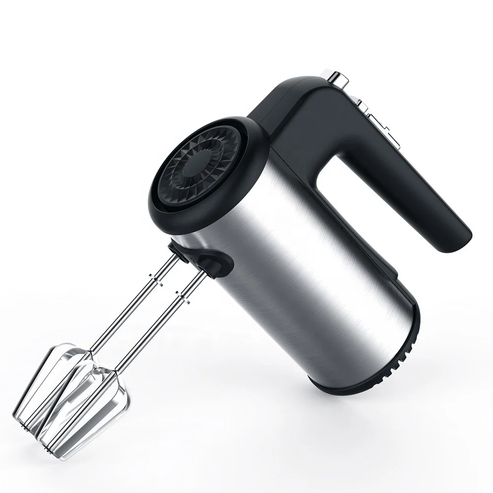
300 Вт безопасный для посудомоечной машины 5-скоростной турбо с крючками для теста, венчик, электрический ручной миксер
571,11-666,30 ₽
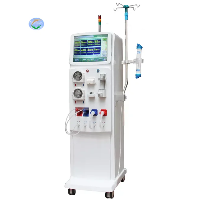
Медицинский портативный прибор для гемодиализа катетер и кровообращения диализа почек ухода за здоровьем
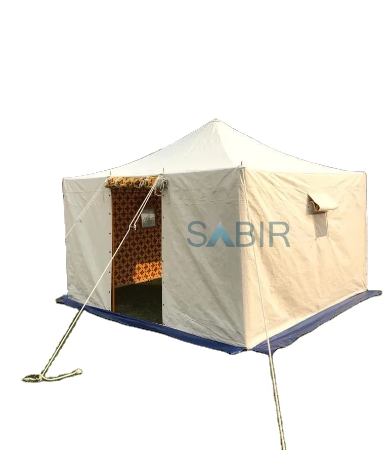
Многофункциональная роскошная водонепроницаемая палатка из брезента для кемпинга однокомнатная всесезонная с настраиваемым размером новый стиль рынка Ближнего Востока
45 688,76-56 159,10 ₽
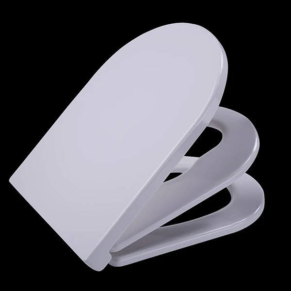
Санитарно-гигиеническое сиденье для
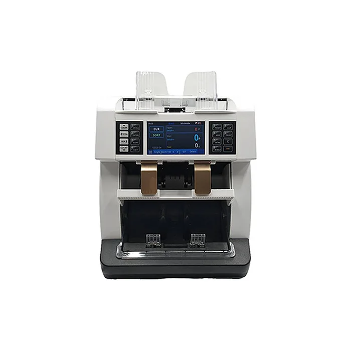
Счетчик купюр с низким уровнем шума на тонкопленочных транзисторах, автоматическое определение разных валют, 4,2 дюйма
50 448-52 351,70 ₽
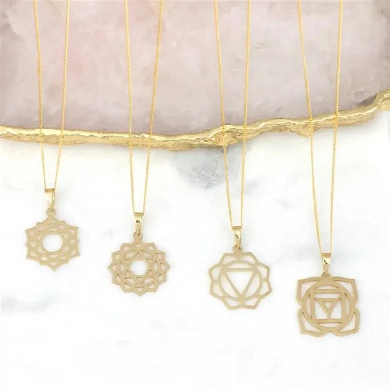
Ожерелье из нержавеющей стали изящное ожерелье с чакрами на заказ модное позолоченное ювелирное изделие водонепроницаемой подвеской без
121,84-217,03 ₽
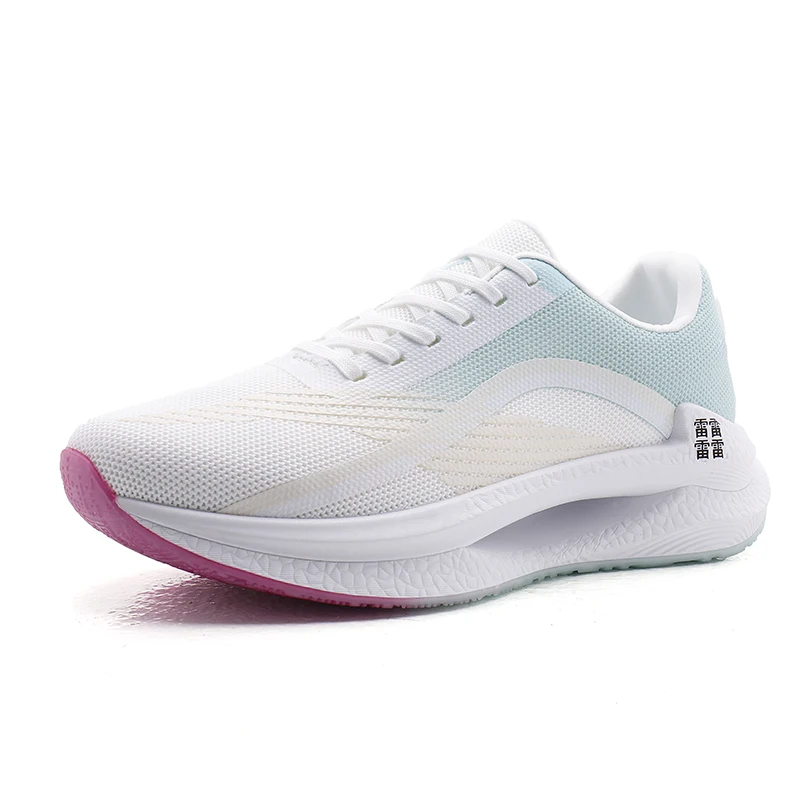
Мужские спортивные кроссовки с логотипом
856,67-999,45 ₽
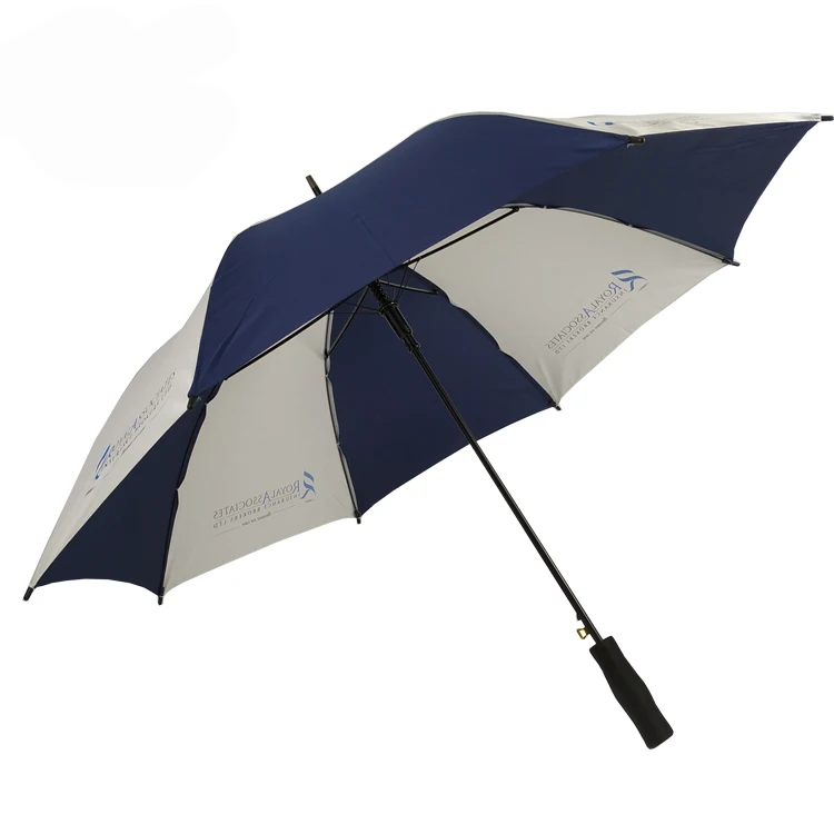
OEM ODM, настоящее изобретение, персонализированный прямой автоматический открытый пользовательский логотип, рекламный зонт для гольфа с логотипом для отелей
842,39-938,53 ₽