Microbiology Prepared Microscope Slides with Specimen used Medicine Education
- Category: Test Instruments >>>
- Supplier: Henan Weike Xiwen Industrial Co. Ltd.
Share on (1600380531680):
Product Overview
Description
Product Description
Zoology Microscope Slide
Since the microorganism is a colorless translucent body, after our technician carefully stained, the morphological structure can be observed clearly. As microorganisms have a relatively constant morphology and structure under certain environmental conditions.Therefore, the study of microbial morphological structure is of great significance in the identification of different groups of
microorganisms, microbiological diagnosis, and analysis of the pathogenicity of pathogens. The observation of microbial morphological structure is an important basic technology in microbiology experiments.
microorganisms, microbiological diagnosis, and analysis of the pathogenicity of pathogens. The observation of microbial morphological structure is an important basic technology in microbiology experiments.

As a manufacturer of prepared slides, we provide OEM service
1, we can prepared slides based your list
2, We have more than 3000 kinds prepared slides, ask us for the list
3, Slides label, your logo is acceptable and different color label
4, Packing box, plastic and wooden material, free to choose
Product details
A total of 21 kinds of dental microscope slides,
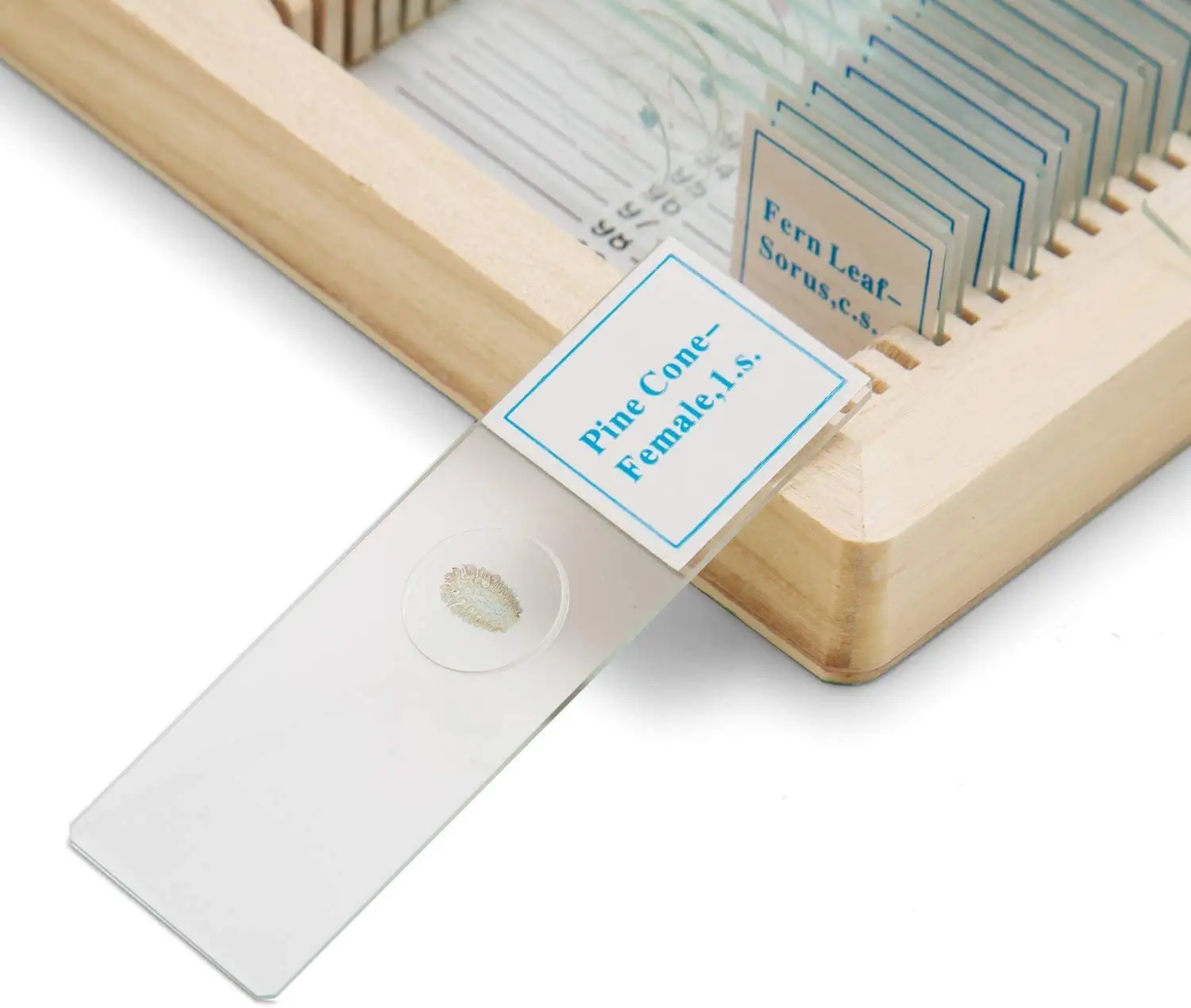
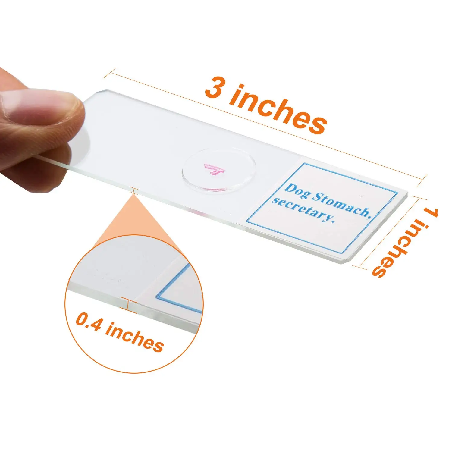
Highly transparent slides, no impurities, to observe the specimen clearly.Manufactured under ISO 9001 quality control standard.
Product security
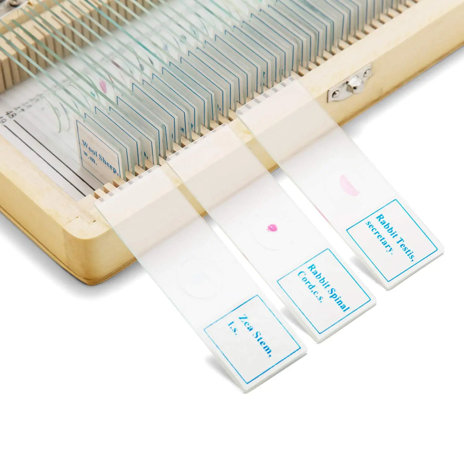
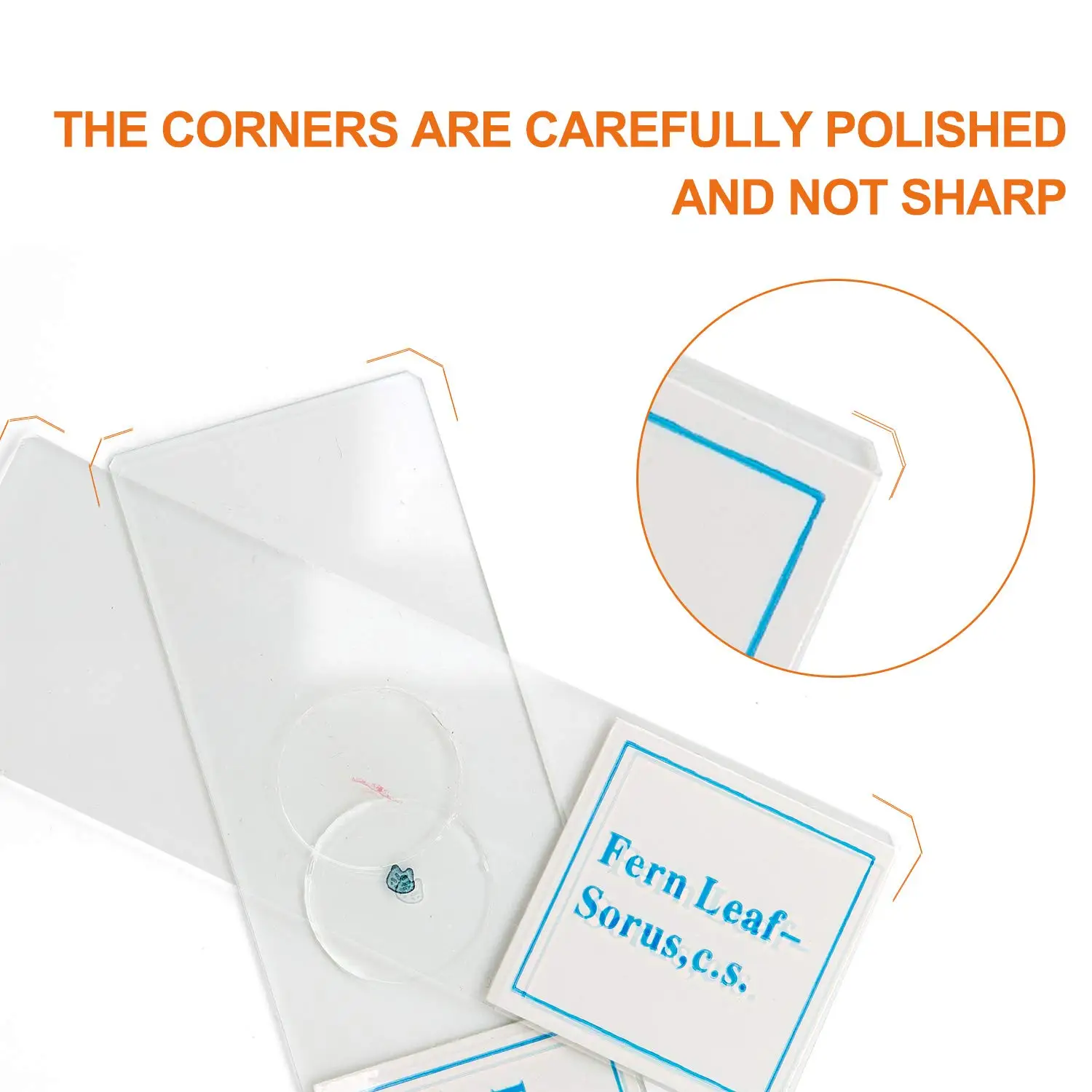
Edge-reinforcing, the slide is made of a separate slot which can be fixed to prevent scratches or breakage.
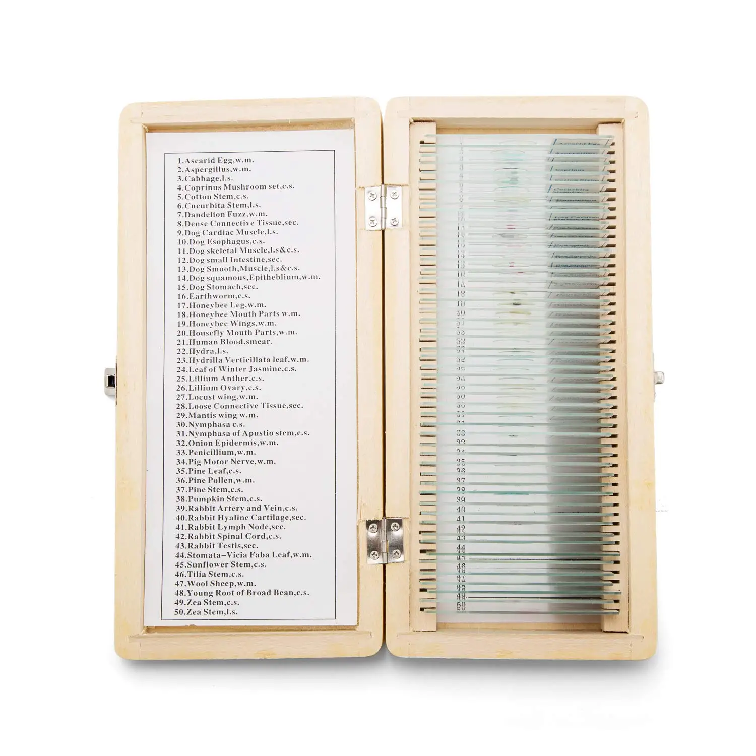
The slides have information labels on one side, making it easy to find the one you're looking for.
The slides have information labels on one side, making it easy to find the one you're looking for.

Specification
We have more than 3000 kinds of different slides include botany, zoology, histology, pathology,parasitology, microbiology, and hematology, etc. Feel free to ask us for our full list or send us your list, you will have our quotation within 12 hours.
01 Euglena w.m. | 51 Climbing leg of louse w.m. |
02 Rotifera w.m. | 52 Membranous wing of honeybee w.m. |
03 Paramecium caudatum w.m. | 53 Coriacrious wing of grasshopper w.m. |
04 Conjugation of paramecium caudatum w.m. | 54 Wing of housefly w.m. |
05 Binary fission of paramecium caudatum w.m. | 55 Lepidotic of butterfly w.m. |
06 Freshwater spongia w.m. | 56 Setaceous antenna of dragonfly w.m. |
07 Spincule of spongia (silica) w.m. | 57 Filiform antenna of grasshopper w.m. |
08 Hydra with bud w.m. | 58 Moniliform antenna of termite w.m. |
09 Hydra t.s. | 59 Serrate antenna of beetle w.m. |
10 Hydra l.s. | 60 Melolontha, cockchafer, antenna with sense organs w.m |
11 Dugesia japanica (intestine injected) w.m. | 61 Geniculate antenna of honeybee w.m. |
12 Dugesia japanica (show reproduction system) w.m. | 62 Aristate antenna of housefly w.m. |
13 Dugesia japanica sagittal section | 63 Whorled antenna of male mosquito w.m. |
14 Adult clonorchis sinensis cobbold w.m. | 64 Clavate antenna of butterfly w.m. |
15 Eggs of clonorchis sinensis cobbold w.m. (nature color) | 65 Drosophila melanogaster w.m. female |
16 Schistosoma japonicum (female) w.m. | 66 Drosophila melanogaster w.m. male |
17 Schistosoma japonicum (male) w.m. | 67 Leg of housefly w.m. |
18 Sexual mosaic of schistosoma japonicum w.m. | 68 The sting of a bee w.m. |
19 Eggs of schistosoma japonicum w.m. (natural color) | 69 Grasshopper testis sec. |
20 Miracidium of schistosoma japonicum w.m. | 70 Grasshopper muscle w.m. |
21 Cercaria of schistosoma japonicum w.m. | 71 Skin of fish |
22 Ascaris lumbricoides (male ) t.s. | 72 Gill of fish t.s. |
23 Ascaris lumbricoides (female) t.s. | 73 Intestine of fish t.s. |
24 Ascaris lumbricoides (female and male) t.s. | 74 Spleen of fish t.s. |
25 Ascarid lumbricoide egg w.m. (natural color) | 75 Blood of fish smear |
26 Hirudo t.s. | 76 Esophagus of fish sec. |
27 Earthworm x.s. | 77 Wall of stomach of fish sec. |
28 Earthworm front median l.s. | 78 Heart of fish t.s. |
29 Earthworm x.s. through clitellum | 79 Fish scale w.m. |
30 Gill of anodonta t.s. | 80 Simple-cell of egg of frog sec. |
31 Daphnia w.m. | 81 2-cell of egg of frog sec. |
32 Cyclops w.m. | 82 Tadpole l.s. |
33 Tetranychus cinnabarinus w.m. | 83 Chromatophores of skin of frog w.m. |
34 Eggs of culex w.m. | 84 Skin of frog t.s. |
35 Pupa of culex w.m. | 85 Liver of frog t.s. |
36 Larva of culex w.m. | 86 Blood of frog smear |
37 Female culex w.m. | 87 Lung of frog t.s. |
38 Male culex w.m. | 88 Ovary of frog sec. |
39 Pediculus humanus w.m. | 89 Intestine of frog t.s. |
40 Young Shrimp entire w.m. | 90 Feather of bird w.m. |
41 Chewing mouthpart of cockroach w.m. | 91 Lung of bird t.s. |
42 Chewing-lapping mouthpart of honeybee w.m. | 92 Gizzard of duck t.s. |
43 Piercing-sucking mouthpart of mosquito w.m. | 93 Ovary of chicken t.s. |
44 Sponging mouthpart of housefly w.m. | 94 Glandulose stomach of chicken t.s. |
45 Siphoning mouthpart of butterfly w.m. | 95 Lung of chicken t.s. |
46 Walking leg of cockroach w.m. | 96 Kidney of chicken t.s. |
47 Grasping leg of mantis w.m. | 97 Tail of mouse (show many tissues) t.s. |
48 Digging leg of mole cricket w.m. | 98 Ovary of cat sec. |
49 Pollen-carrying leg of honeybee w.m. | 99 Esophagus of dog t.s. |
50 Jumping leg of grasshopper w.m. | 100 Trachea of dog t.s. |
WM(W.M): Whole Mount(Entire Specimen or Organism) LS(L.S): Longitudinal Section. A section cut lengthwise.Cut parallel to the longitudinal axis.
CS(C.S): Cross-Section.Such as a thin wafer of an Earthworm.Cut perpendicular to the longitudinal axis.
TS(T.S): Transverse.An alternative name for cross-section.
Sec : Section Sm/Smear:Smear Sq:Squashed preparation.
CS(C.S): Cross-Section.Such as a thin wafer of an Earthworm.Cut perpendicular to the longitudinal axis.
TS(T.S): Transverse.An alternative name for cross-section.
Sec : Section Sm/Smear:Smear Sq:Squashed preparation.
FAQ
▶What is the difference between biological slices, mounts, and smears?
Biology is an experimental natural science. The main content of biology is the knowledge of the form, structure, and physiology of various organisms in nature. When students learn about the internal structure and microstructure of organisms, it is not easy to understand. If they can display vivid specimens in front of students, the problem will naturally be solved.
Slice: A flat thin part cut from an object, such as a leaf vein slice.
Mounting tablets: Mounted tablets taken from living organisms or directly made from individual tiny organisms, such as onion epidermal cells and human oral cavity cells.
Smear: A specimen made by applying a liquid specimen (such as blood, bone marrow fluid, etc.) collected from a living organism evenly on a glass slide, such as a human blood smear.
▶How are the products packaged?
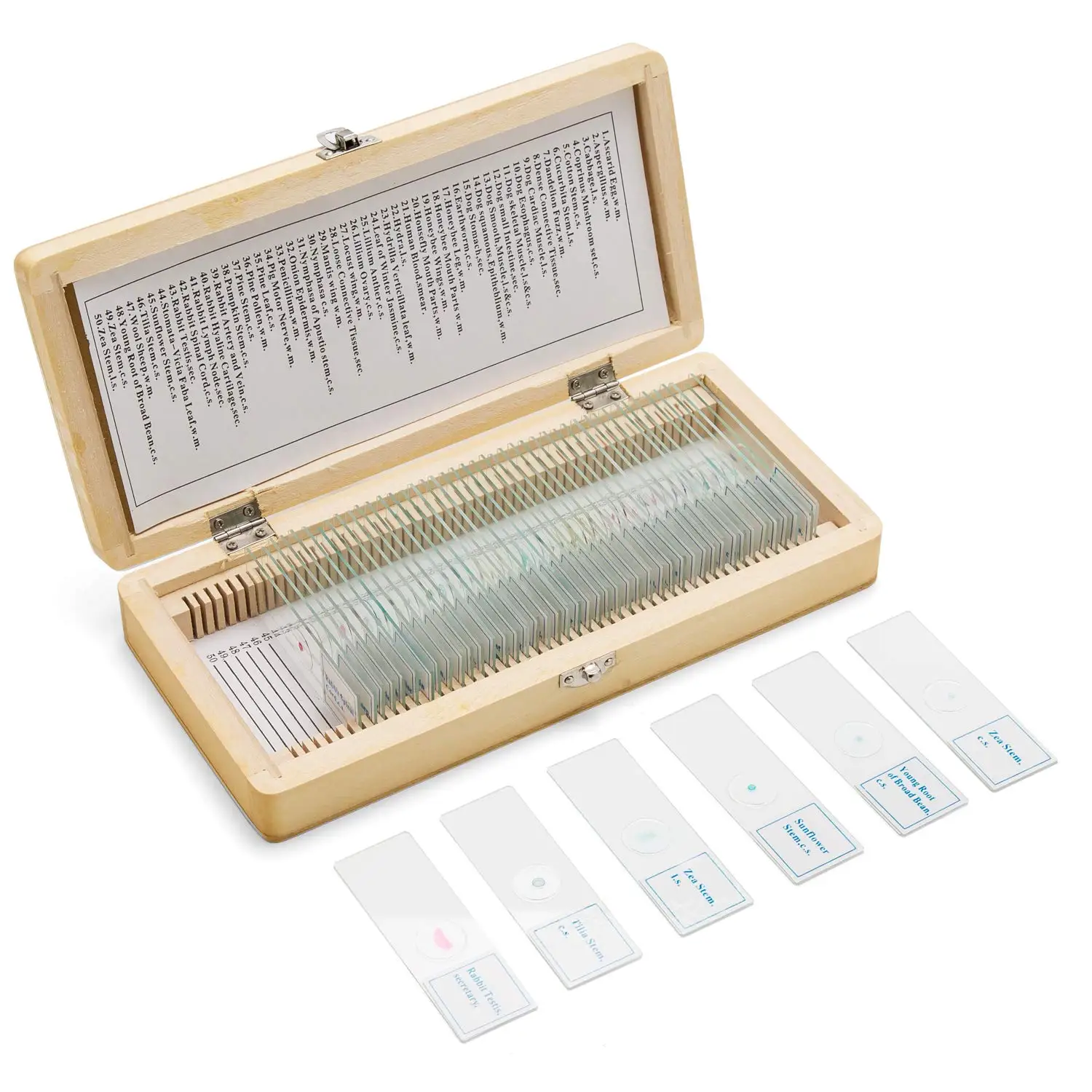
This product has a total of two kinds of packaging box, plastic packaging box and wooden packaging box. Plastic box has a built-in sponge protection to prevent any damage during transportation or movements, and which effectively saves the product,the number of slide coding inside is clear at a glance,convenient to take. Wooden box has all wood structure, fine workmanship, beautiful and practical, conducive to long-term preservation of biological slides,Metal clasp lock is exquisite and stable, not easy to oxidize,the slot is even, and the slide is arranged neatly and fixed, to prevent friction or breakage.
▶What is your minimum order quantity?
The minimum order quantity is 1 piece, the more the quantity, the more preferential price.
Slice: A flat thin part cut from an object, such as a leaf vein slice.
Mounting tablets: Mounted tablets taken from living organisms or directly made from individual tiny organisms, such as onion epidermal cells and human oral cavity cells.
Smear: A specimen made by applying a liquid specimen (such as blood, bone marrow fluid, etc.) collected from a living organism evenly on a glass slide, such as a human blood smear.
▶How are the products packaged?
This product has a total of two kinds of packaging box, plastic packaging box and wooden packaging box. Plastic box has a built-in sponge protection to prevent any damage during transportation or movements, and which effectively saves the product,the number of slide coding inside is clear at a glance,convenient to take. Wooden box has all wood structure, fine workmanship, beautiful and practical, conducive to long-term preservation of biological slides,Metal clasp lock is exquisite and stable, not easy to oxidize,the slot is even, and the slide is arranged neatly and fixed, to prevent friction or breakage.
▶What is your minimum order quantity?
The minimum order quantity is 1 piece, the more the quantity, the more preferential price.

Packing & Delivery
Packing instructions: This product is packed in wooden boxes and plastic boxes, and shock-proof sponge is used inside the box to prevent damage during transportation or movement.The outside of the box is made of explosion-proof foam and tightly packed with tape to provide multiple protection barriers for the product.
Product Process
This shop specializes in customizing various biological specimen slices,including the different usage categories of teacher demonstration and student experiment.Very representative, the varieties cover botany, zoology,microbiology,parasitology,histology, and human histology. It is a good choice for students to explore the microscopic world and stimulate interest using microscopes.

Company Profile
Henan Weike Xiwen Industrial Co.,Ltd
Henan Weike international trade Co.,Ltd has become the country’s largest professional biological microslides、specimen production import and export,provincial high-tech incubator key enterprise、biological microslides in national colleges of higher learning、recoomended brand of specimen、import and export company domestic designated biological microslides processing and export enterprises,the company has passed IS9001;2000 quantity management system certification.Biological microscope slides include:botany, zoology, microbiology, tissue pregnancy,Cell biology and genetics, animal pathology, plant histopathology, human histology, etc.Originated from education, service education, technological innovation, and integrity management.It is precisely because of our excellent corporate team, high-quality products, and superior after-sales service that the company's continuous development has been achieved.We satisfy users with strong strength and continuous research and development of new products,we sincerely welcomes you and we will serve you wholeheartedly.


We Recommend


New Arrivals
New products from manufacturers at wholesale prices